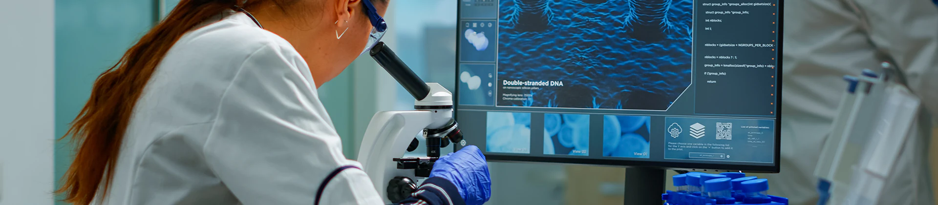
Electron Microscopy
Our service unit houses a high-resolution Field Emission Scanning Electron Microscope (FESEM). The FESEM, which uses a field emission electron gun as its electron source, is a type of scanning electron microscope that enables the examination of surface details at the nanometer scale. Compared to conventional SEM, it produces much finer and denser electron beams, allowing for high-resolution imaging.
Instruments and Equipment
- Zeiss Sigma 500, Electron Microscope (FESEM)
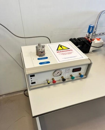
- Quorum K850, Critical Point Dryer
- Quorum Q150R S, Coating System
- Leica EM TRIM2, Epon Block Preparation System
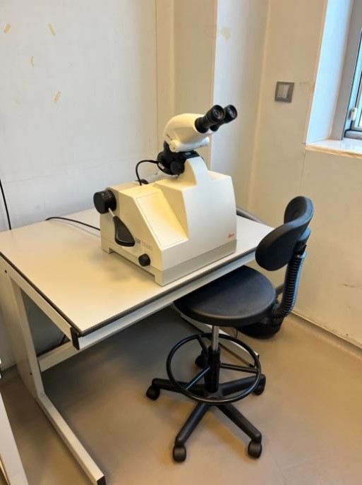
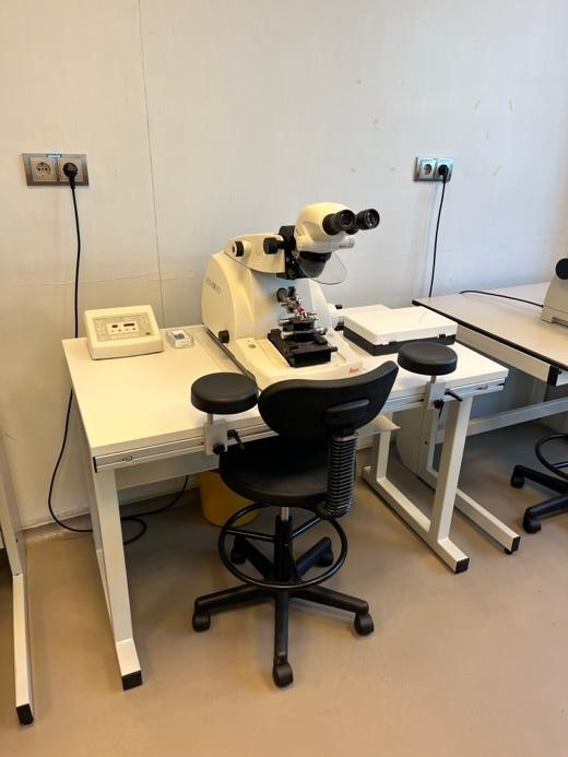
- Leica EM UC7, Ultramicrotome
Services
1- SEM Sample Preparation
In our unit, SEM sample preparation is performed using the Quorum K850 critical point dryer, which allows biological specimens, polymeric, and various material samples to be dried and made ready for imaging. Additionally, for non-conductive samples, the Quorum Q150R S coating system enables coating with gold/palladium (Au/Pd), making them suitable for SEM analysis.
 |
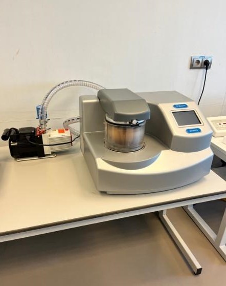 |
| Quorum K850 Critical Point Dryer | Quorum Q150R S Coater |
2- TEM Sample Preparation
For TEM sample preparation, following fixation and tissue processing, Epon blocks can be trimmed using the Leica EM TRIM2 system. Semi-thin and ultra-thin sections can then be obtained with the Leica EM UC7 ultramicrotome available in our laboratory.
 |
 |
| Leica EM TRIM2 |
Leica EM UC7 Ultramicrotome |
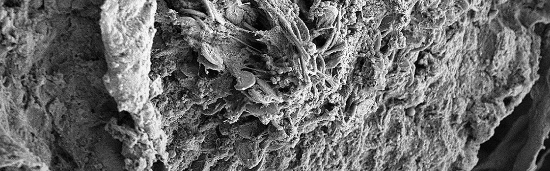
3- STEM/SEM Imaging
The Zeiss Sigma 500 electron microscope in our center operates within a 20 V – 30 kV range (0.8 nm at 15 kV, 1.6 nm at 1 kV). With its integrated EDS detector, the quantitative distribution of elements within a sample can be determined, and elemental mapping can be performed. Using the EBSD detector, the crystallographic structure of the sample can be analyzed in detail, while the STEM detector allows for high-resolution examination of biological specimens, thin films, and polymeric materials.

Zeiss Sigma 500 (FESEM)
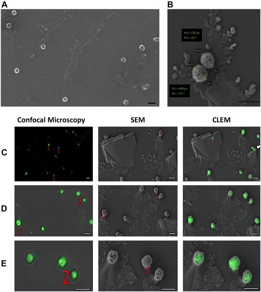
Additionally, our unit provides Correlative Microscopy studies. In this approach, a cell or material is first observed with a light microscope in its fluorescently labeled state, and the same region is then analyzed in detail with the electron microscope for structural characterization.
Reference:
Demir Ş, Erdal E, Bagriyanik HA. Imaging of Isolated Exosomes by Correlative Microscopy. Journal of Histochemistry & Cytochemistry. 2024;72(3):149-156. doi:10.1369/00221554241233346
Unit Coordinator: Gökçen BİLİCİ GÜLER