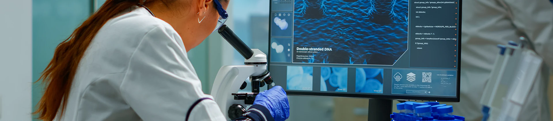
Optical Imaging
Optical imaging plays a critical role in numerous research areas, including monitoring intracellular dynamics, analyzing protein interactions, studying cell migration, morphological changes, and tracking signaling pathways. With its advanced microscopy systems and expert technical staff, the Optical Imaging Unit supports a wide range of scientific projects, from basic research to preclinical studies.
The unit has the capacity to image a broad variety of biological samples—from live cells to tissue sections—with high resolution. Thanks to its advanced technological infrastructure, it provides services to both internal and external researchers, enabling the visualization of biological processes. Throughout the imaging workflow, users are supported in experiment design, sample preparation, imaging parameter optimization, and image analysis.
Instruments
- Zeiss LSM880, Confocal Microscope
- Zeiss Observer 7, Apotome Fluorescence Microscope
- Olympus BX61, Fully Motorized Fluorescence Microscope System
- Olympus IX71, Fluorescence Microscope
- Olympus CKX41, Light Microscope
- Olympus IX81, Live Cell Imaging Microscope
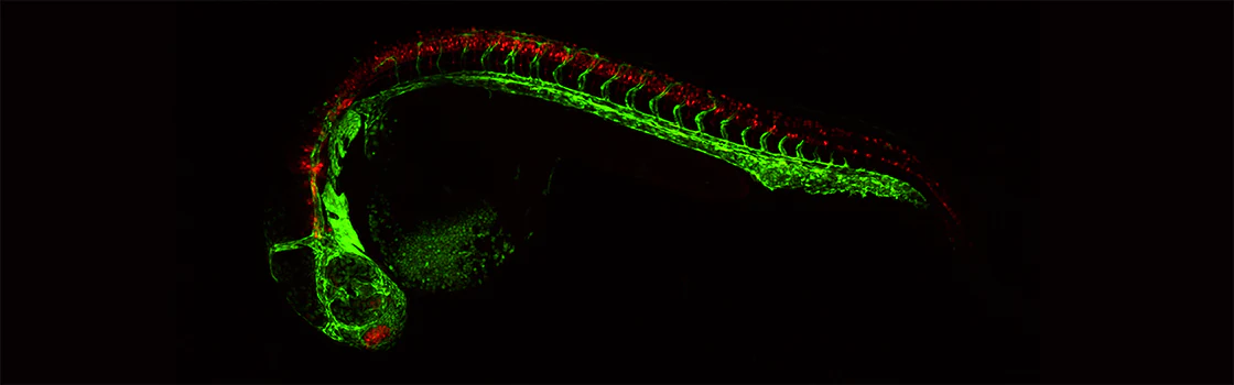
With these instruments, advanced techniques can be applied, including multichannel fluorescence imaging, 2D imaging, 3D imaging (Z-stack), tile scan, time series (capturing images at specific times and locations to generate videos), and super-resolution imaging (Airyscan).
Additionally, the Zeiss LSM880 confocal microscope enables correlative light and electron microscopy (CLEM) analyses in integration with SEM.
Services
- High-resolution imaging of fixed and live cells
- Detailed cellular structure analysis via confocal microscopy
- Long-term live cell monitoring and time-lapse imaging
- Image analysis support using ImageJ, ZEN, and other software
- Consulting for cell counting, co-localization, time series analysis, and related applications
- Guidance on experimental design and protocol development
Unit Coordinator: Aslı SEREN